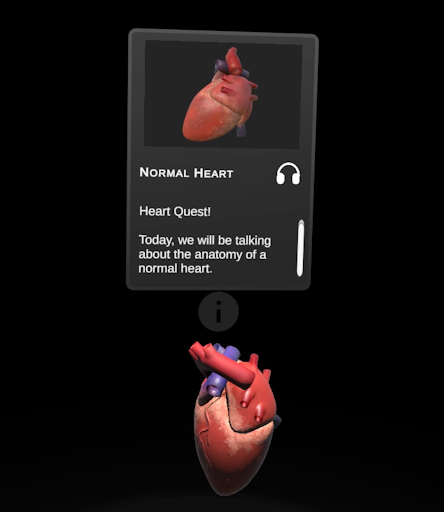
Last summer, Pediatric Cardiologist Dr. Jason Mendel and Cardiology Fellow Dr. Hope Kile reached out to Studio X about creating an educational VR application focused on congenital heart disease (CHD). They wanted to help people better visualize and understand congenital heart defects through 3D models.
Existing applications and anatomical models didn’t quite meet their needs. Some were inaccessible to the public or behind academic and commercial paywalls. Others were medically accurate but difficult for someone without a medical background to understand. They wanted something in between: anatomically accurate models that the general public could access and understand.
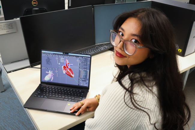
I’m Aadi Rajbhandary. When I worked on HeartQuest, I was a senior computer science major at the University of Rochester and the project’s 3D modeling leade. I came to the project with experience in VR development and 3D modeling, but almost no background in anatomy or medicine. Before beginning, I couldn’t have confidently identified the different parts of the heart, let alone the defects I would eventually model. Hope and Jason brought the clinical expertise to understand what each defect looked like and which details mattered. Much of our work became a joint effort to translate between two very different areas of expertise.
Before our first meeting, Hope and Jason shared examples of existing applications, including the Stanford Virtual Heart Experience. We discussed what they wanted from the experience and its tentative scope, but there was still a big gap between the idea and what the final application would look like. It took experimentation and trial and error to bridge it.
As we developed the storyboard and clarified what we wanted URochester HeartQuest to accomplish, our early conversations focused on the experience itself: Who was it for? What should someone understand after taking off the headset? How much medical knowledge could we assume they already had?
We also asked what VR could add to traditional educational resources. Congenital heart defects change complex three-dimensional structures, and their spatial relationships can be difficult to understand from a two-dimensional diagram. In VR, users could physically explore those differences: pick up a heart, rotate and scale it, open it to examine its internal anatomy, and view the defect from different perspectives. We wanted people to actively explore the anatomy rather than passively receive information.
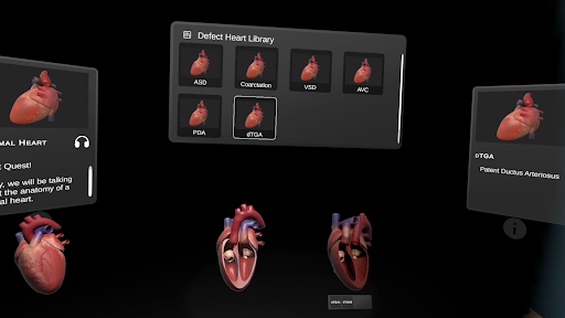
That idea shaped the structure of HeartQuest. For each defect, we decided to show three models side by side: a standard heart, a heart with the selected defect, and a heart after surgical correction. Users can move between them to compare the anatomy, first learning the structure of a standard heart, then seeing what has changed, and finally examining how surgery addresses that change. We also incorporated audio explanations so users could listen while exploring the models.
We had to be realistic about the project’s scope, given the timeline and my academic and Studio X responsibilities. We prioritized interactions that directly supported the educational goals. Scaling, rotating, and opening the hearts offered different ways to examine the anatomy, while the side-by-side presentation gave users a consistent way to compare defects.
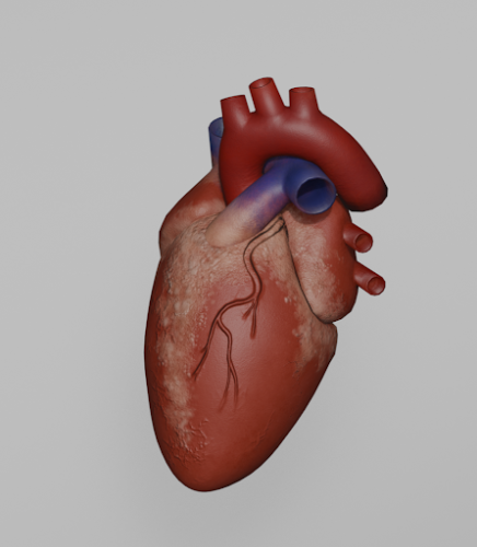
Defining these interactions early also clarified what the models needed to do. They had to look accurate from the outside and hold up when users enlarged them, viewed them from different angles, or opened them to see internal structures. First, though, I needed to understand and build the standard heart that would serve as the foundation for the other models. My Karp Mentor, Emmie, worked with me on that initial model.
One of the biggest hurdles was finding reference images that gave me enough information to model the heart. A medically accurate image or scan wasn’t always easy for me to interpret. I might receive a diagram and realize I didn’t know which structure I was looking at or how structures shown in two dimensions connected in three.
As I became more comfortable modeling the heart, I learned that making something look anatomically convincing is very different from making it anatomically correct. I was used to asking whether a shape matched its reference, whether the proportions looked right, and whether the model made sense from different angles. But a structure could look convincing and still connect in the wrong place, follow the wrong path, or misrepresent an important feature.
This became especially clear as I moved from the standard heart to individual defects and their surgical repairs. Small details could distinguish one condition from another, so I couldn’t rely on my interpretation alone to decide when a model was finished. Each model needed review by Hope and Jason, and their feedback became essential to the process.
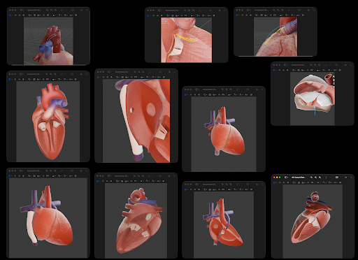
At first, the feedback process was informal. I interpreted the references, created the model in Blender, and sent screenshots for review. But screenshots didn’t always show a 3D model well, especially when we needed to discuss internal structures or how different parts related to one another. An issue might only become clear when we met and could rotate the model or point directly to a structure.
This created a longer feedback loop than I expected. I might complete a section based on my interpretation, only to learn at our next meeting that I had misunderstood an underlying structure. I also didn’t always know which views would help Hope and Jason assess the anatomy. Their medical perspective and my 3D modeling perspective made some corrections difficult to describe asynchronously. Finding time to meet around academic, Studio X, and clinical schedules could also take a while.
We adapted as we went. Instead of finishing one model before getting feedback, I began creating initial versions of several defects at once, focusing first on their major shapes and structures. I shared videos as well as screenshots so Hope and Jason could see the models from all sides, and we scheduled check-ins to review several models together.
This made the work more efficient. I could keep moving on other models while waiting for clarification, and we could validate the major anatomy before I spent time refining details, texturing, and optimizing each model for VR. That reduced the chance of reworking a model I had already considered finished.
I also got better at asking for feedback. Early on, I didn’t always know enough anatomy to identify what confused me, so my questions were often as broad as, “Does this look right?” As I learned more, I began asking about specific connections, positions, or internal structures. Pinpointing what I didn’t understand made our conversations more productive and helped me incorporate the medical feedback directly into the models.
The models also had to work as part of an interactive VR experience. They needed to hold up when users picked them up, rotated them, scaled them, and examined them from different angles. I had to consider not only whether the anatomy was correct, but whether users could understand it in VR.
One challenge was deciding how to open each heart. Many defects are inside the heart and can be hard to see from the outside. Cutting every model in the same place wouldn’t necessarily expose the anatomy most relevant to that defect. I experimented with different cuts, trying to reveal important structures while keeping enough of the surrounding anatomy to show where the defect was located.
One approach I started calling the “cake cut” removes a wedge-shaped section, like taking a slice out of a cake. It gives users a view inside while leaving most of the heart’s shape intact, helping them understand where a defect sits in relation to the rest of the anatomy. The cut creates a “big slice” and a “small slice,” offering two possible views. Only the small slice is in the experience now, but the larger one may be useful for future models.
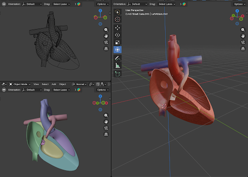
I also had to balance anatomical detail with VR performance and efficient texturing. I reduced unnecessary geometry while preserving structures that helped explain each defect. Whenever possible, I kept the standard heart’s geometry consistent across the defect and surgical repair models, changing only what was needed. This let me reuse textures and maintain a consistent look across the experience.
One of the biggest things I learned from HeartQuest was how to work on a project where I was not the subject expert. At first, that was intimidating. I knew how to model and build for VR, but not enough about the heart to make decisions about anatomical accuracy. Over time, I realized I didn’t need to become a medical expert. My role was to learn enough to understand what Hope and Jason were communicating, ask questions when I didn’t, and translate that information into something I could build in 3D.
I also became more comfortable admitting when I didn’t understand something. Early on, I sometimes spent too long trying to interpret a reference on my own because I wasn’t sure what to ask. As I learned the anatomy, I got better at noticing those moments and saying, “I understand this part, but I don’t understand what is happening here.” Asking a good question could be more useful than trying to find an answer alone.
The project changed how I think about iteration, too. We revised the models, but we also adjusted how we worked together. When screenshots weren’t enough, we used videos and met more often. When completing one model at a time led to too much rework, I changed my approach. A lot of HeartQuest came together by recognizing what wasn’t working, changing it, and trying again.
HeartQuest now includes 20 models representing seven congenital heart defects and their surgical repairs. With the initial development complete, the next stage will focus on evaluating how effectively the models communicate the anatomy they represent. We also plan to make the models publicly available as open educational resources and eventually publish the complete experience.
When I began working on HeartQuest, I couldn’t have confidently identified most of the structures I would spend months modeling. By the end, I had a much stronger understanding of cardiac anatomy and had learned how to design in a field where I wasn’t the expert. To me, designing across disciplines means learning where your expertise ends and someone else’s begins, then building a shared process that brings those perspectives together. HeartQuest could not have been created through medical expertise or 3D modeling expertise alone. It took both, working toward the same goal.
About this Author
Aadi Rajbahindary, XR Specialist & & 3D Modeling Team Lead